| FIG. 2 |
|
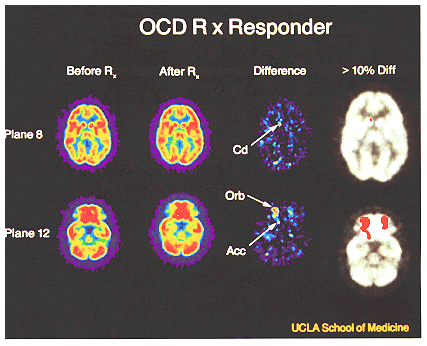
| Subject with severe OCD studied with FDG-PET for glucose metabolic rates before and after effective treatment with paroxetine hydrochloride. PET image plane 8 is at the level of the head of the caudate nucleus, whereas plane 12 is at the level of the orbital gyri and nucleus accumbens. The first two columns (left to right) show normalized glucose metabolic rate images before and after 6 weeks of paroxetine treatment. Difference column shows subtraction of posttreatment from pretreatment image to show decreases in glucose metabolic rates. The last column shows difference data windowed to show regions of significant (>10%) decreases in glucose metabolic rate, in red, and superimposed on image of underlying neuroanatomy in the same patient. These PET results seen after decreasing OCD symptoms with drug treatment can be viewed as the inverse of those seen in Fig. 1, where OCD symptoms were increased with provocation. |
| Back to Chapter |
published 2000