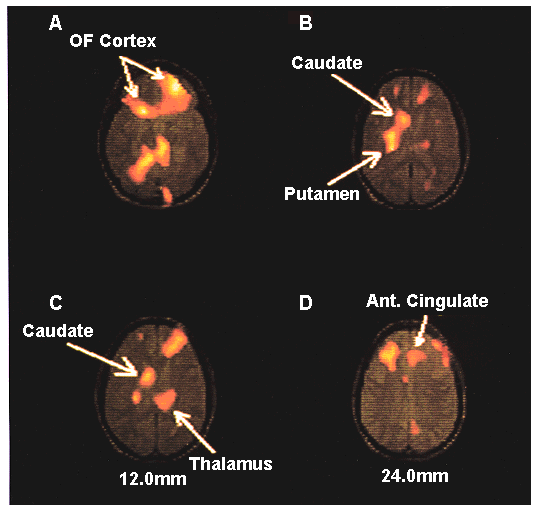
PET study measuring cerebral blood flow rates in eight OCD subjects exposed to a neutral stimulus, then with one designed to provoke obsessive thoughts and anxiety. Shown are omnibus subtraction images (summed for all subjects) with the provoked state minus the resting condition, displayed with a hot-iron scale in units of z scores, superimposed over a normal magnetic resonance image transformed to a standard brain, for the purpose of anatomic reference. Increasing z score indicates activity in the provoked state that is increased to levels above that in the resting state.
Orbital cortex, bilaterally, and right head of caudate nucleus were a priori brain regions of interest, which showed significant (p < 0.01) changes (increases); other regions were significant at the trend level (p < 0.10). From Rausch et al. (60).