| Figure 5. |
|
|
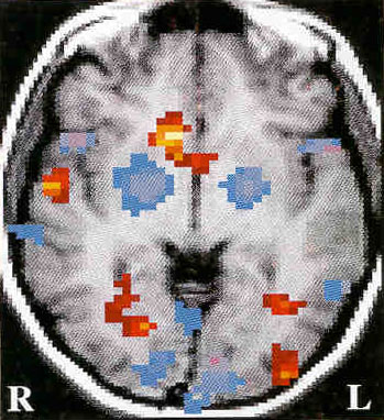
| Voluntary Tic Suppression during a Functional Magnetic Resonance Imaging Study. Representative single-subject activation map during voluntary tic suppression obtained during a study of 22 adult TS subjects showing bilaterally decreased activity in the putamen and globus pallidus (blue) and increased activity in the right caudate nucleus (red). Periods of voluntary tic suppression were compared with periods when subjects were tic as freely as possible given the constraints of the MRI scanner. Magnitudes of the signal changes were correlated with tic severity outside the scanner (R putamen, Pearson r= 0.54, p<.01; L putamen, Pearson r = 0.40, p=.07; and R caudate, Pearson r = 0.46, p<.02. For further details see ref. 84. |
published 2000