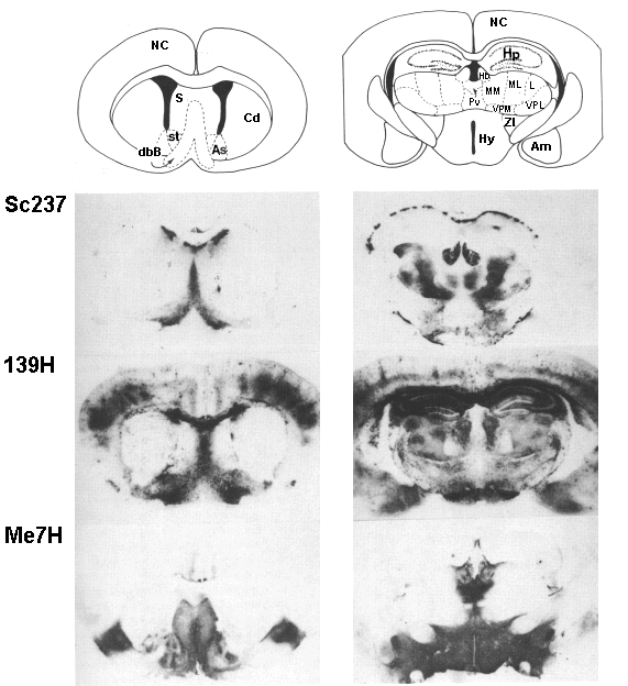
FIG. 2

|
FIG. 2 |
 |
| The distribution of PrPSc in the brain of Tg(SHaPrP)-7 mice which express high levels of Syrian hamster PrPC is unique for the Sc237, 139H, and Me7H prion isolates (19). Histoblots from two levels of the brain are compared for each prion isolate. The location of PrPSc was obtained by pressing 10-mm cryostat sections to nitrocellulose paper, digesting the resulting histoblot with proteinase K to eliminate PrPC, and denaturing the remaining protease resistant PrPSc with quanidinium to enhance antibody binding followed by immunohistological localization with PrP-specific antibodies. |
| Back to Chapter |
published 2000