| FIG. 1 |
|
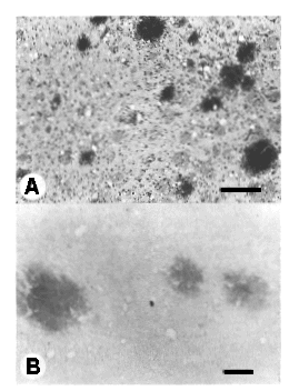
| PrP cerebral amyloid plaques develop in the spontaneous neurodegenerative disease of transgenic mice which express a mouse PrP gene mimicking the codon 102 mutation genetically linked to human ataxic GSS, Tg (MoPrP-P102L)-174 mice (Hsiao et al., in preparation). Most of the amyloid plaques were located in the caudate nucleus but also occurred in the hippocampus and cerebellar cortex. A: Multiple amyloid plaques in the caudate nucleus. Bar represents 100 mm. Periodic acidSchiff stain. B: Amyloid plaques in the caudate nucleus react specifically with PrP antibodies (R073). Peroxidase immunohistochemistry following proteinase K digestion. Bar represents 50 mm. |
| Back to Chapter |
published 2000