| Figure 1 |
|
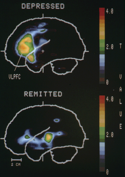
| Increased activity in the left prefrontal cortex was found in depressed but not the remitted subjects. Each t-image indicates areas where subjects in the depressed (top) and remitted (bottom) phases of familial pure depressive disease have increased tissue radioactivity relative to the control subjects. Mean voxel t-values for the sagittal planes from 4751 mm left of the bicommissural line are demonstrated. With permission from Drevets et al. 1992 (25). |
| Back to Chapter |
published 2000