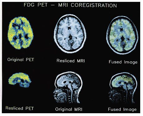
| Figure 4. |
|
|
| Reslicing and co-registration of structural MRI and FDG PET images of glucose metabolism from a 39 year old healthy female control. Original MRI was acquired in the sagittal plane, and original FDG PET in the axial plane. Surface matching (Analyze) software enabled images in each modality to be resliced to match the orientation of the other modality, and then fused to provide anatomic definition for functional image. The PET scan was obtained on a 7 slice Scanditronix PC1024-7B dedicated head scanner with an in-plane resolution of 5.2mm and an axial resolution of 11mm. The tracer was 5mCi of FDG. There were 4 transmission scans (one for each frame) with a rotating Ge/Ga pin source. The subject had a hexalite head holder and eyepatches on and performed an auditory continuous performance task for 30 minutes and was then scanned for 30 minutes (4 frames of 7 slices to yield 28 interleaved slices). The interslice distance is 13.75mm for the 7 original slices and hence 3.4375mm for the 28 slices resulting from the 4 interleaved frames. The PET image resliced in the sagittal plane clearly shows the brainstem, cerebellum and fourth ventricle. The sagittally acquired MRI was obtained on a Picker 0.5T scanner. Slice thickness 2.5mm, acquired with 2 repetitions. 256 x 256 pixels x 64 slices. It is a T1 weighted field echo scan with TR=36, TE=6, flip angle=30 degrees. Figure provided courtesy of T. Ketter, NIH. |
published 2000