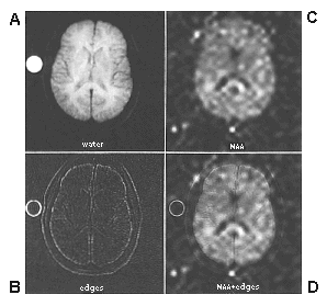
| Figure 3. |
|
|
| Co-registration of structural MRI and proton MRS images, acquired in the same scan session from a healthy young control. Left column (upper) illustrates unprocessed axial water-based structural image, and (lower) edge-detected version. Right column (upper) illustrates proton inversion recovery MRS image of n-acetylaspartic acid (NAA) distribution in the axial plane, and (lower) a linear combination of NAA distribution and structural edges providing neuroanatomic definition without obscuring spectroscopic data. Figure provided courtesy of Daniel Spielman, Department of Radiology, Stanford University. |
published 2000