| Figure 3 |
|
|
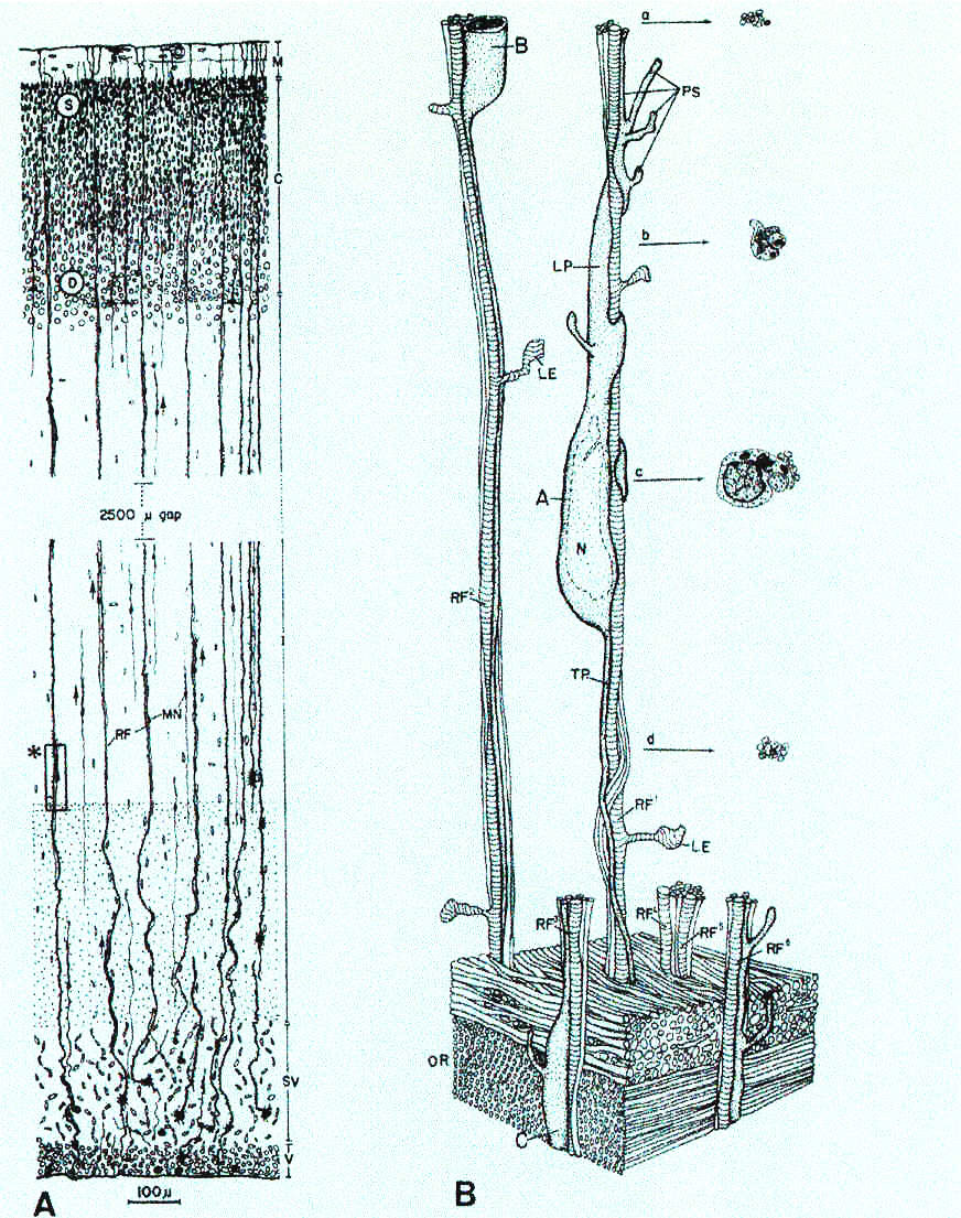
| Mode of cell migration to the primate cerebral cortex. Left, camera lucida drawing of the occipital cerebral wall of the monkey fetus at midgestation. The rectangle marked with an asterisk shows the approximate position of the cell reconstruction shown in B. Right, three-dimensional reconstruction of migrating neurons. Except at the lower portion of the figure, the fiber system has been omitted to expose the radial fibers (RF) and their relationship to the migrating cells A,B and C. Migrating cell A is shown with its nucleus (N), leading process (LP) and vertical ascending pseudopodium (PS); cross-sections of A at different levels are shown at the right side of the figure. Abbreviations: C, cortical plate; I, intermediate zone; M, molecular layer; MN, migrating neuron; RF, radial fiber; SV, subventricular zone; V, ventricular zone; OR, optic radiation; S, superficial and D, deep layers of the cerebral cortex. From Rakic (1972) , reproduced with permission. |
published 2000