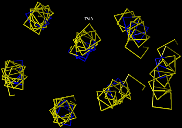
The spatial orientation of highly conserved residues (blue) in a rhodopsin-based 5-HT2A receptor model. The model is shown as an extracellular view of a trace of a-carbons only. The helices are ordered in a counter-clockwise fashion.
| Figure 8. |
|
|
|
The spatial orientation of highly conserved residues (blue) in a rhodopsin-based 5-HT2A receptor model. The model is shown as an extracellular view of a trace of a-carbons only. The helices are ordered in a counter-clockwise fashion. |
| Back to Chapter |
published 2000