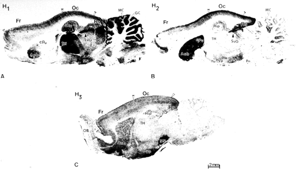
Autoradiographic localization of histamine receptors on midsagittal sections of brain. H1 and H2 receptors were visualized on sections of guinea pig brain using [125I]iodobolpyramine and [125I]iodoaminopotentidine, respectively (A,B). H3 receptors were visualized on section of rat brain using [125I]iodoproxyfan (C). Abbreviations: Acb, nucleus accumbens; cPu, caudate putamen; Fr, frontal cortex; GC, granular layer of cerebellum; Hip, hippocampus; Hyp, hypothalamus; MC, molecular layer of cerebellum; OB, olfactory bulb; Oc, occipital cortex; Pn, pontine nuclei; Rs, retrosplenial cortex; SN, substantia nigra; SuG, superficial gray layer of superior colliculus; TH, thalamus; Tu, olfactory tubercle; VTg, ventral tegmental nucleus.