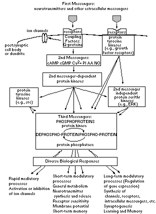
Schematic illustration of the role played by intracellular messenger systems in synaptic transmission in the brain. Recent studies in neuroscience have provided a complex view of synaptic transmission. These studies have focused on the involvement of intracellular messenger systems, involving coupling factors (termed G- proteins), second messengers [e.g., cAMP, cGMP, Ca2+, nitric oxide (NO), and the metabolites of phosphatidylinositol (PI) and arachidonic acid (AA)], and protein phosphorylation (involving the phosphorylation of phosphoproteins by protein kinases and their dephosphorylation by protein phosphatases), in mediating multiple actions of neurotransmitters on their target neurons.
Prominent in brain are numerous protein serine/threonine kinases that are activated directly by various second messengers, and are referred to as second messenger-dependent protein kinases. Brain also contains numerous protein serine/threonine kinases that are not regulated directly by second messengers (see Table 1). In addition, the brain contains numerous types of protein tyrosine kinases, some of which reside in the receptors for most growth factors, others that are not associated with growth factor receptors (see Table 1). These various protein kinases are all highly regulated by extracellular stimuli. The second messenger-dependent protein kinases are regulated by receptor-second messenger pathways as shown in Figure 3. The receptor-associated protein tyrosine kinases are activated upon growth factor binding to the receptor. The second messenger-independent protein serine/threonine kinases and the protein tyrosine kinases that are not receptor-associated seem to be regulated indirectly by second messengers and second messenger-dependent protein kinases and by protein tyrosine kinases, although the precise mechanisms are not yet known in most cases. The brain also contains numerous types of protein serine/threonine and protein tyrosine phosphatases, which are also subject to regulation by extracellular stimuli.
The figure illustrates three major roles subserved by these intracellular messenger pathways. In some cases, intracellular messengers mediate the actions of some neurotransmitters in opening or inhibiting particular ion channels. However, intracellular messengers mediate most of the many other actions of neurotransmitters on their target neurons. Some are relatively short-lived and involve modulation of the general metabolic state of the neurons, their ability to synthesize or release neurotransmitter, and the functional sensitivity of their various receptors and ion channels to various synaptic inputs. Others are relatively long-lived and are achieved through the regulation of gene expression in the target neurons. Thus, neurotransmitters, through the regulation of intracellular messenger pathways and alterations in gene transcription and protein synthesis, alter the numbers and types of receptors and ion channels in target neurons, the functional activity of the intracellular messenger systems in those neurons, and even the shape and numbers of synapses the neurons form. The figure is drawn to illustrate the amplification that intracellular messenger systems can give to neurotransmitter action. Thus, a single event of a neurotransmitter binding to its receptor (the 1st messenger level) can act through the 2nd, 3rd, 4th, etc. messenger levels to produce an increasingly wider array of physiological effects.