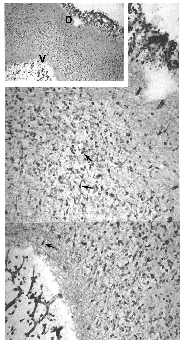
| Figure 2. |
|
|
| The low-power insert illustrates the region of the mesencephalic flexure through its full dorsal (D) to ventral (V) extent. This section is adjacent to the midline; therefore the aqueduct of the mesencephalon is not visible. Nevertheless, newly born DA neuroblasts migrate from the germinal epithelium ventrally, and in so doing they provide the appearance seen in the high-power photomontage of small, bipolar neurons with elongated neurites that extend in a dorsoventral plane. The migrating neurons (arrows) are stained for tyrosine hydroxylase and demonstrate the early onset of transmitter enzyme in these neurons as depicted at E 4041. |
published 2000