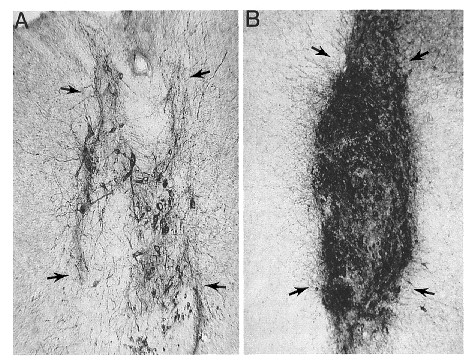
| Figure 10. |
|
|
| Representative transplants (arrows) of embryonic DA neurons are seen in two adult African green monkeys at the same magnification. This comparison of grafted neurons collected from an optimally aged donor (B) versus those of an older donor (A) illustrates the dramatic increase in viability seen following grafting of the younger tissue. These illustrations are representative of grafts that contain approximately 500 (older donor) and 5000 (younger donor) tyrosine-hydroxylase-positive neurons as reflected by the density of perikarya. This enhanced survival undoubtedly relates to the immature stage of neuritic development at the earlier time, but may be attributed to other factors as well. |
published 2000