| Figure 5. |
 |
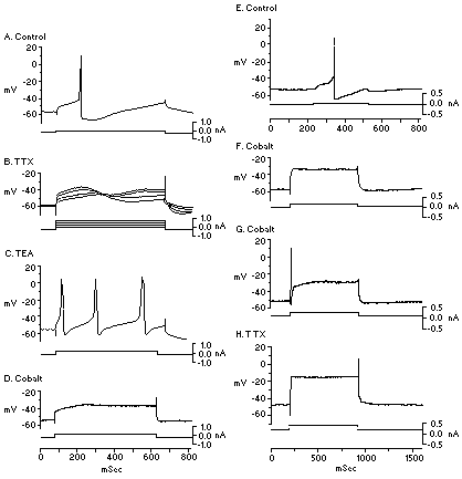
| Action potentials recorded in vitro in DA neurons can be differentiated pharmacologically into two components. A: Injection of a depolarizing pulse (bottom trace) evokes a membrane depolarization and action potential discharge (top trace) in an identified DA neuron. B: After the administration of 1 mm of the sodium channel blocker tetrodotoxin (TTX) into the superfusion fluid, the depolarization-evoked spike discharge is blocked even when the amplitude of the membrane depolarization is increased severalfold. C: Administration of the selective potassium channel blocker tetraethylammonium (TEA; 2 mM) enables moderate amplitudes of membrane depolarization to elicit a series of large-amplitude, long-duration spikes with prominent spike afterhyperpolarizations. D: Subsequent application of the calcium blocker cobalt (2 mM) prevents the occurrence of these depolarization-elicited high-threshold spikes (HTSs). Other experiments show that this HTS underlies the somatodendritic component of the action potential. E: Depolarization of the membrane of a DA neuron in control conditions evokes action potential discharge. F: Administration of the calcium blocker cobalt to the superfusion fluid blocks spike activity in this neuron. G: However, if sufficient amplitudes of membrane depolarization are delivered, the cell discharges a moderate-amplitude, fast spike. This spike is brief in duration and is associated with very little spike afterhyperpolarization. H: Subsequent administration of the sodium channel blocker TTX blocks this depolarization-elicited spike discharge. Other experiments show that this TTX-sensitive spike underlies the initial segment spike of the action potential. (From ref. 17, with permission.) |
| Back to Chapter |
published 2000