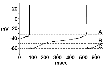
| Figure 3. |
 |
| DA neurons recorded intracellularly in vivo and in vitro exhibit characteristic pacemaker-like slow depolarizations and high spike thresholds. In this recording from a spontaneously discharging DA neuron recorded in vitro from a rat midbrain slice, a slow membrane depolarizing conductance depolarizes this neuron from its resting potential (B) to the atypically high membrane potential threshold for spike generation that is characteristic for these neurons, which in this case is -33 mV (A). The action potential is followed by a calcium-dependent afterhyperpolarization (C), which then decays prior to the initiation of a subsequent slow depolarization. (From ref. 29, with permission.) |
| Back to Chapter |
published 2000