| Figure 3. |
 |
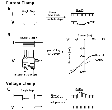
| Schematic of typical protocols for current- and voltage-clamp recording. A: Current-clamp. Left panel: Membrane potential is recorded with a brief injection (20–500 msec) of a rectangular negative (hyperpolarizing) current (I) pulse through the recording barrel; a hyperpolarizing voltage (V) response (deflection) is recorded that reflects the impedance (resistive) properties of the membrane (effects of electrode resistance are subtracted). The slowness of the onset and offset of the voltage response represents the effect of membrane capacitance and resistance. The amplitude of the voltage response is proportional to membrane resistance; this value times the current injected allow the calculation of "input resistance" (by Ohm's law). Right panel: During injection of multiple current pulses at regular intervals (2–10 sec) and recording at a slower time-base, voltage responses appear as brief downward deflections. Application of a conventional inhibitory transmitter such as GABA reveals the expected hyperpolarization associated with a reduction of the size of the downward deflections, indicating reduced input resistance. B: To allow better analysis of voltage-dependent drug effects (see Fig. 4) and to determine their reversal or equilibrium potentials, construction of voltage–current curves are required. Here, multiple pulses or steps of current of both polarities and various intensities (e.g., 0.05–2 nA) are injected at regular intervals and resultant voltage responses displayed superimposed at high oscilloscope speed, as shown at left. The "sag" in the voltage responses often seen at more hyperpolarized potentials results from a time-dependent current [e.g., the M-current (see Fig. 4) or the Q or h current, a hyperpolarization-dependent inward rectifying current involved in pacemaker potentials]. Plotting the size of these potentials (at either the peak or the steady state: dotted lines) against the current (the independent variable) yields a V–I curve (right panel) with properties characteristic for each cell. Repeating this procedure during drug application may then reveal changes in the position and slope of the V–I curve. For example, GABA lowers the curve and reduces the slope, indicating hyperpolarization with reduced input or slope resistance (or increased conductance). The intersection of the control and GABA curves at around -80 mV denotes the reversal potential of the GABA effect, as would be expected of increased Cl- conductance and recording with a pipette filled with an anion that does not alter the intracellular Cl- concentration. C: A simple voltage-clamp protocol, in which the membrane current is the measured, dependent variable. Voltage is the independent variable: Using a single hyperpolarizing voltage command (Vc), with the membrane potential clamped (at holding potential: Vh, often around resting potential), one can obtain a relative estimate of the "macroscopic" current (left panel) flowing through multiple ion channels (see text). The slow time-dependent inward "relaxation" in the current trace during the command potential represents the so-called Q or h current. When multiple voltage commands of the same size are applied repetitively at slow chart speed (right panel), one can determine whether a drug increases or decreases ionic conductance (directly proportional to the size of each current response); thus, in this case, GABA increases ionic conductance. Not shown: Delivery of multiple depolarizing and/or hyperpolarizing voltage commands of different intensities can be used to generate current–voltage curves similar (although with axis rotated 90 degrees and the curve inverted) to the V–I curves of current-clamp recordings (see part B, right panel). Such I–V curves give an estimate of "slope conductance" of the macroscopic currents over a large membrane potential range. Here, increased slope would indicate increased conductance (decreased resistance). (Modified from Fig. 7 of ref. 5.) |
published 2000