| Figure 5. |
 |
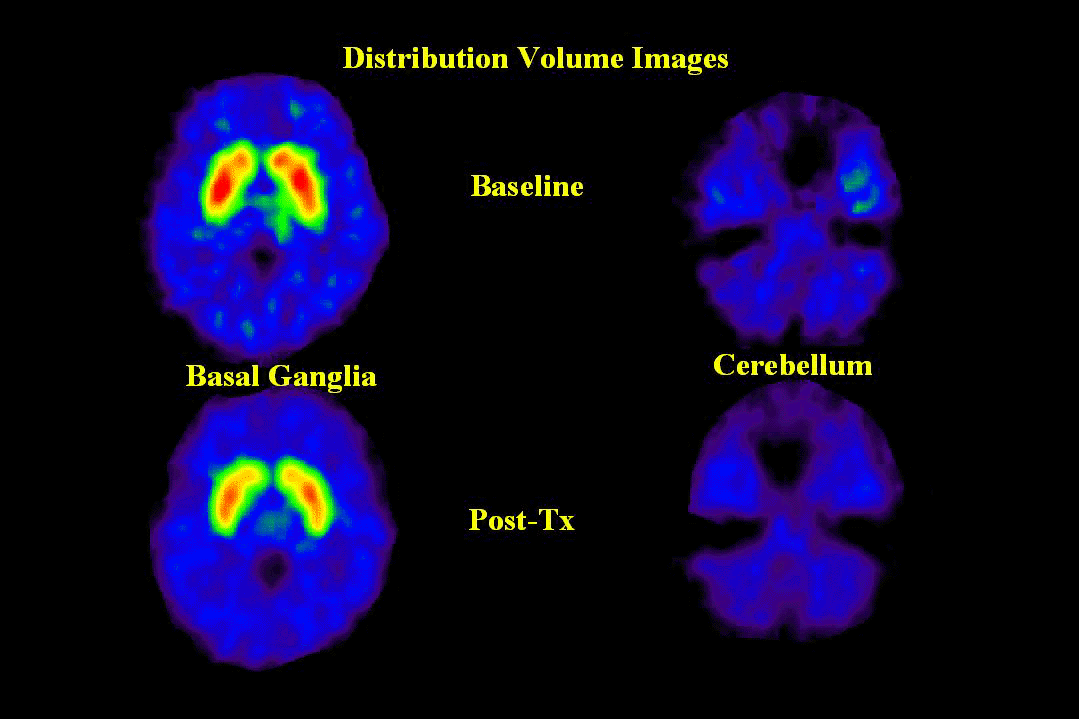
| PET images of [11C]-raclopride binding from a representative sujbect are shown at the level of the basal ganglia (left) and cerebellum (right). The scans in the upper panel were performed under baseline conditions and those in the lower panel were performed three hours after fenfluramine administration (60 mg, po). Note the decrease in [11C]-raclopride binding after fenfluramine administration in the basal ganglia (specific binding) and the lack of change in the cerebellum (non-specific binding). |
published 2000