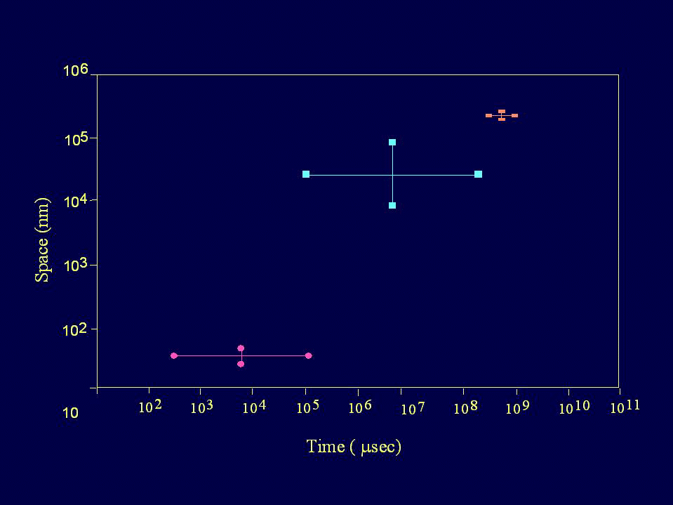
| Figure 3. |
|
|
| A representation of the relation between the dynamics of transmitter release at the synaptic cleft (l ) and the in vivo sampling methods of voltammetry (¨ ) and microdialysis (n ) with respect to the temporal and spatial domains. Calculations are based on a synaptic cleft of 30 - 50 nm with a synaptic delay of 0.3 - 100 msec, voltammetric electrodes of 10- to 100-μm diameter with a sampling interval of 100 msec to 3 min, and microdialysis probes of 250- to 300-μm diameter and a sampling interval of 5 - 15 min. |
| Back to Chapter |
published 2000