| Figure 8. |
|
|
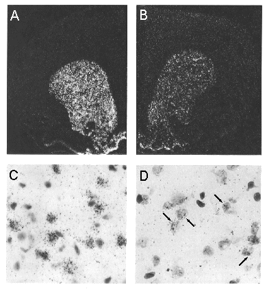
| Dark-field photomicrographs in A and B illustrate distribution pattern of neurons expressing mRNA transcripts encoding the D2 dopamine receptor in the rat forebrain as revealed by in situ hybridization. The riboprobe used in the experiment shown in A was directed against an exonic portion of the D2 mRNA, whereas the probe applied in the experiment in B was specific for an intronic segment of the D2 hnRNA. Cellular labeling from the two cases is seen at higher magnification in the bright-field photomicrographs in C and D, which were from sections that were dipped in emulsion and Nissl-counterstained. Note the cytoplasmic distribution of grains from the experiment involving the exonic probe C, in contrast to the largely nuclear distribution of grains following application of the intronic probe D. (Courtesy of Dr. Charles A. Fox and Dr. Alfred Mansour.) |
published 2000